Written By
Ari Magill, MD, BS
Updated on May 28, 2024
Litigation Guides
MCA Strokes
The middle cerebral artery (MCA) is the brain's largest artery and the most common site for strokes. Strokes in this area can cause severe symptoms, including loss of movement, speech, and awareness of one side of the body. Acute treatments include IV medication to dissolve clots or mechanical devices to remove them. Stroke prevention typically involves taking aspirin or similar medications and managing risk factors.
Written By
Ari Magill, MD, BS
What is an MCA Stroke?
The middle cerebral artery (MCA) is the brain's largest artery and the most frequent location for strokes, a medical emergency where the blood supply to part of the brain is interrupted, causing brain cells to die and leading to potential disability or death.1,2,3
The MCA has four main branches (M1, M2, M3, M4) supplying blood to crucial brain regions.1,2,3 These regions include:1,2
- Most of the outer brain surface, including the:
- Frontal lobe - the brain's control center for decision-making, behavior, and voluntary movements
- Temporal lobe - part of the brain’s surgace, located around the temples, that processes sound, language, and memory
- Parietal lobe - a part of the brain’s surface that processes touch, spatial awareness, and body sensations
- Deeper structures like the:
- Caudate - a brain structure involved in motor control and learning
- Internal capsule - a brain structure that carries information between different areas of the brain and spinal cord
- Thalamus - a central brain relay station that processes and sends sensory and motor signals to the rest of the brain

Stroke Symptoms
The extensive area supplied by the MCA translates to a wide range of potential symptoms depending on the affected branches and structures.2 Large MCA strokes are generally easier to diagnose compared to other stroke types due to their often-pronounced symptoms. Common symptoms include:2
- Weakness on one side of the body
- Forced eye movement in one direction
- Loss of vision in specific areas of the visual field
- Difficulty with spoken language (understanding, producing words, or forming sentences) if the dominant brain hemisphere (usually left) is affected.
MCA strokes can cause various impairments beyond the typical ones. These may include:1
- Neglect (inattention to one side of the body)
- Lack of coordination (ataxia)
- Changes in perception
- Bowel and bladder problems
- Cognitive issues
- Swallowing difficulties
- Subtle communication and vision problems
The brain's left hemisphere is dominant for language functions (sentence formation, word structure, pronunciation).4 Damage to the left hemisphere can cause aphasia, a language communication difficulty. In contrast, the right hemisphere is more involved in:4
- Spatial awareness (hemispatial neglect: ignoring one side of the body/environment)
- Processing the "big picture" rather than details
- Providing context for information and categories
Strokes affecting specific areas, particularly the non-dominant (usually right) hemisphere, can cause:5
- Confusion
- Agitation
- Restlessness
Likewise, vertebrobasilar ischemia (reduced blood flow to the back of the brain) can lead to sudden confusion with memory loss, mimicking delirium.5 A key factor in diagnosis is the acute onset - unlike other causes of delirium that typically develop more gradually.
How are MCA Strokes Diagnosed?
Clinical history and neurological examination remain the foundation for diagnosing ischemic strokes caused by reduced blood flow.6 Several factors can complicate diagnosing MCA strokes:7
- Incomplete initial information available to healthcare providers
- Symptoms that evolve over time
- Variability in brain regions supplied by different arterial branches, leading to atypical presentations
- Small strokes, early stroke stages, young stroke patients, and strokes affecting posterior (back) brain regions may present with less obvious deficits. This can make diagnosis based solely on clinical signs difficult.
Modern neuroimaging techniques have significantly improved the ability to:6
- Locate brain damage.
- Identify blood clot locations within and outside the brain.
Stroke mimics
Certain conditions can mimic strokes, leading to initial diagnostic challenges. Examples of stroke mimics include:5,6
- Low blood sugar (hypoglycemia)
- Atypical migraine headache
- Seizure-related paralysis (postictal paralysis)
- Rapidly developing brain tumors
- Brain infections (abscesses, or pockets of pus, and encephalitis, or inflammation of the brain)
- Nerve fiber damage (demyelination)
- Bell's palsy (facial paralysis due to peripheral nerve injury)
How to recognize stroke mimics
Diagnosing stroke mimics requires thorough investigation, often including MRI scans when necessary. These conditions are more prevalent in:5
- Younger patients
- Women
- Individuals with milder symptoms compared to typical stroke presentation
Notably, up to one-third of stroke mimic patients may not exhibit any detectable neurological deficits during examination.5 Additionally, research suggests stroke mimics tend to have normal or lower blood pressure readings.
Diagnostic challenges in stroke
Common symptoms like loss of consciousness, vomiting, and headache can occur in both stroke mimics and real strokes.5 This overlap in symptoms makes definitive diagnosis based solely on these factors difficult.
Vertebrobasilar stroke, affecting the back of the brain and brainstem, can also present with minimal or no neurological abnormalities.5 Patients with this type of stroke often report nausea, vomiting, imbalance, or dizziness.
Acute neuroimaging
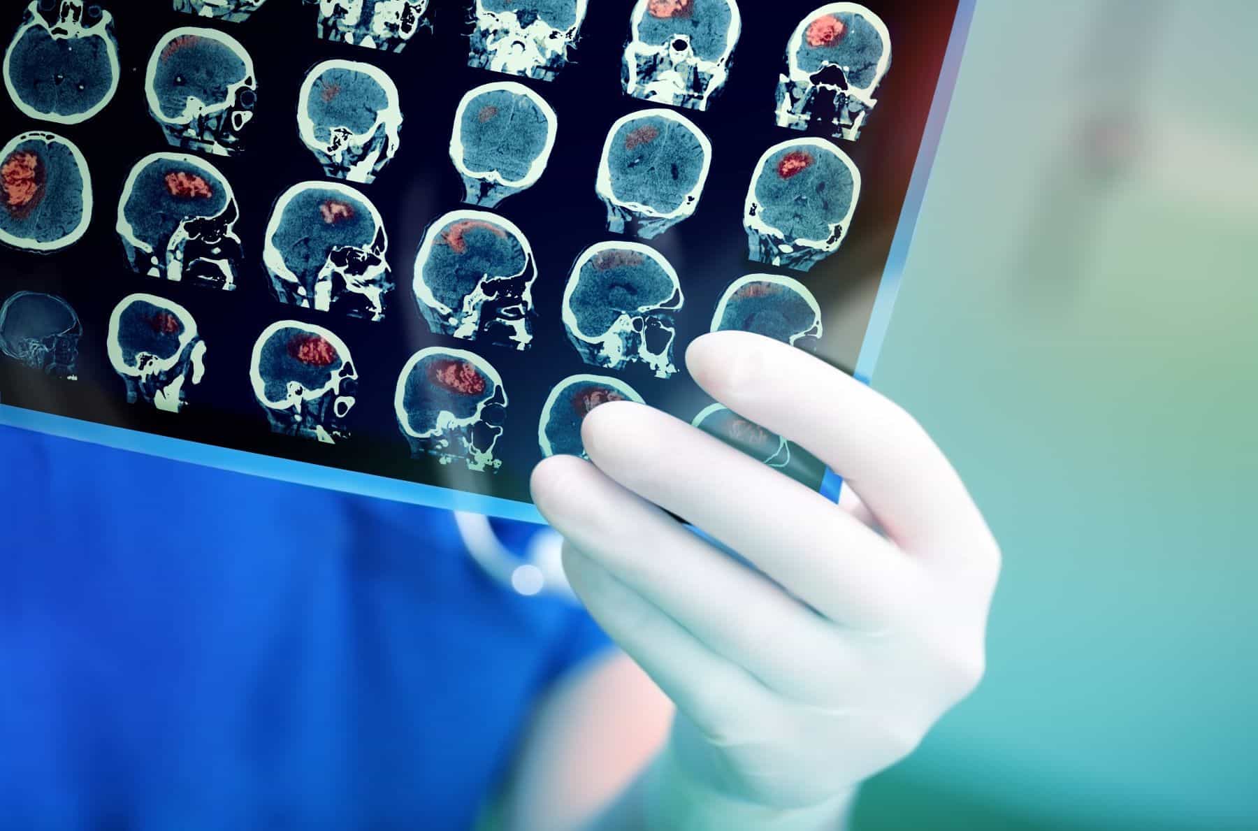
Computed tomography (CT) scan findings are primarily used to rule out bleeding, tumors, and other non-stroke lesions. For example:6
- Acute bleeding appears as a dense mass.
- Tumors and infections appear as mass lesions (growth of abnormal cells) with varying densities.
- Ischemic strokes may show subtle changes initially, especially in deep brain regions.
MRI diffusion-weighted imaging (DWI) is:5
- Highly accurate for detecting recent strokes within minutes of symptoms.
- Can differentiate stroke from mimics, but limitations exist.
Key takeaways
When the diagnosis is unclear, remember:8
- Sudden neurological deficits suggest stroke until proven otherwise.
- Gradual symptom onset, inconsistent exams, or atypical symptom patterns may indicate stroke mimics.
- Neurological consultation is crucial in unclear cases.
Navigating a MCA Stroke Case?
We help attorneys access the latest legal research, medical record reviews, physician consultations, and world-class experts.
Stroke Prevention
Lifestyle changes can help prevent stroke recurrence. These include:9
- regular exercise
- maintaining a normal weight
- eating a healthy, high-fiber diet with plenty of vegetables and fruits
- managing blood pressure
- quitting smoking
- addressing known risk factors
Stroke Treatment
The goal of ischemic stroke treatment is to rapidly restore blood flow to the affected brain area.10 Treatments can be divided as follows:
Thrombolysis (clot-busting drugs)
Timing is critical to effective thrombolytic treatment, as described below:6,10
- Imaging and treatment decisions should ideally occur within 45 minutes for potential thrombolysis within a 0-4.5 hour window.
- Benefits increase with earlier administration.
Options for thrombolysis are as follows:10
- Alteplase is the primary clot-dissolving medication used to treat stroke.
- Tenecteplase is an alternative with potentially stronger clot-dissolving abilities and better outcomes, but further research is needed.
Intra-arterial thrombolysis
Intra-arterial thrombolysis is a potential option for acute MCA blockages within 6 hours of stroke onset. It can be further described as follows:10
- Involves delivering clot-dissolving medication directly to the blocked artery.
- May be considered for patients ineligible for systemic thrombolysis or outside the 4.5 hour window.
Mechanical clot removal (thrombectomy)
Key points regarding use of mechanical thrombectomy can be summarized as follows:10
- Mechanical thrombectomy may be needed when clot-dissolving medications do not work or cannot be used.
- For large vessel occlusions like those in the MCA, mechanical thrombectomy is often the most effective treatment, as clot-dissolving medication alone may not be enough.
- A combination of clot-dissolving medication and mechanical clot removal significantly improves the chances of reopening the blocked vessel.
- This combined treatment has been found to be more effective in restoring blood flow and functional independence in patients at 90 days after the stroke.
- The time frame for using mechanical clot removal has been extended from 6 hours to up to 24 hours in certain patients.
To be eligible for this treatment, patients must meet specific criteria:10
- High level of functional ability before the stroke (less than three on the modified Rankin scale, a measure of how much a person's stroke has affected their daily activities and independence).
- High National Institute of Health Stroke Scale (NIHSS) score (indicating greater severity of the stroke).
- Evidence of a large vessel blockage in the front part of the brain (anterior circulation), as shown by CT angiography or MR angiography.
- Potential for brain tissue recovery, as indicated by CT perfusion or DW-MRI sequences, which should reveal only a limited volume of brain tissue already destroyed by the stroke (small infarct core volume).
Conventional medical therapy
Not all MCA stroke patients fit in the inclusion and exclusion criteria for thrombolysis or mechanical clot removal, so many patients only receive conventional medical therapy, which usually consists of:11
- Blood pressure management
- Blood clot prevention
- Supporting recovery
- Addressing other stroke risk factors
Key points regarding antiplatelet therapy (medication to prevent blood cells called platelets from clumping together and forming clots) can be summarized as follows:6
- Aspirin and similar medications can be beneficial within 48 hours of stroke onset.
- Benefits include preventing blood clots, reducing stroke risk, and improving overall outcome.
- However, a 24-hour window is recommended after clot-dissolving medications (rt-PA) to avoid complications.
A recent study examined long-term function in MCA stroke patients receiving only standard medical treatment (no clot-removal procedures) and came to the following conclusions:11
- Better pre-stroke functional abilities (higher NIHSS and Barthel index scores) predicted better long-term outcomes.
- Lower pre-stroke hemoglobin A1c (average blood sugar levels over the past two to three months) and cholesterol levels were also positive prognostic factors.
- Conversely, high cholesterol, chronic kidney disease, and dementia were linked to poorer outcomes.
- Better blood flow through the carotid artery and the absence of coronary heart disease were associated with a greater chance of functional improvement.
Works Cited
1.
Slater DI. Middle Cerebral Artery Stroke: Overview, Rehabilitation Setting Selection and Indications, Best Practices. eMedicine. Published online January 19, 2023. https://emedicine.medscape.com/article/323120-overview#a1
2.
Nogles TE, Galuska MA. Middle Cerebral Artery Stroke. StatPearls [Internet]. Published 2023. https://www.ncbi.nlm.nih.gov/books/NBK556132/
3.
Paradiso S, Anderson BM, Boles Ponto LL, Tranel D, Robinson RG. Altered Neural Activity and Emotions Following Right Middle Cerebral Artery Stroke. Journal of Stroke and Cerebrovascular Diseases. 2011;20(2):94-104. https://www.dropbox.com/scl/fi/3jpowl2p0jd3febb0q8zz/paradiso2011.pdf?rlkey=icm66wkh7rnoieqo4vdlvjx2g&e=1&dl=0
4.
Seikel JA. An Attentional View of Right Hemisphere Dysfunction. Clinical Archives of Communication Disorders. 2018;3(1):76-88. https://e-cacd.org/journal/view.php?doi=10.21849/cacd.2018.00276
5.
Pohl M, Hesszenberger D, Kapus K, et al. Ischemic stroke mimics: A comprehensive review. Journal of Clinical Neuroscience. 2021;93:174-182. https://www.jocn-journal.com/article/S0967-5868(21)00481-1/fulltext
6.
Gorelick PB, Ruland S. Diagnosis and Management of Acute Ischemic Stroke. Disease-a-Month. 2010;56(2):72-100. https://www.jocn-journal.com/article/S0967-5868(21)00481-1/fulltext
7.
Edlow JA, Selim MH. Atypical presentations of acute cerebrovascular syndromes. The Lancet Neurology. 2011;10(6):550-560. https://www.dropbox.com/scl/fi/mhxk8giu1g1wnyzin3klt/1-s2.0-S1474442211700692-main.pdf?rlkey=2o33rr3kfvorwvcfczgw19u0a&e=1&dl=0
8.
Long B, Koyfman A. Clinical Mimics: An Emergency Medicine-Focused Review of Stroke Mimics. The Journal of Emergency Medicine. 2017;52(2):176-183. https://www.dropbox.com/scl/fi/05kgh1cog1mjunncfmfqt/long2016.pdf?rlkey=zfepqrpu7ygc37hjftw5d3lg9&e=1&dl=0
9.
Wang L, Li H, Hao J, et al. Thirty-six months recurrence after acute ischemic stroke among patients with comorbid type 2 diabetes: A nested case-control study. Frontiers in Aging Neuroscience. 2022;14:999568. doi:https://doi.org/10.3389/fnagi.2022.999568
10.
Ganesh RV, Luoma V, Reddy U. Acute management of ischaemic stroke. Anaesthesia & Intensive Care Medicine. 2022;23(12):747-753. https://www.dropbox.com/scl/fi/x0dodky5q5aoswy6fuyua/1-s2.0-S1472029922002375-main.pdf?rlkey=a972cnpewrc12m6wm65zfu8xs&e=1&dl=0
11.
Yang JL, Lin CM, Hsu YL. Long-Term Functionality Prediction for First Time Ischemic Middle Cerebral Artery Stroke Patients Receiving Conventional Medical Treatment. Neuropsychiatric Disease and Treatment. 2022;18:275-288. https://www.dovepress.com/long-term-functionality-prediction-for-first-time-ischemic-middle-cere-peer-reviewed-fulltext-article-NDT
About the author
Subscribe to our newsletter
Join our newsletter to stay up to date on legal news, insights and product updates from Expert Institute.
Speak with a Stroke Expert
We're here to help you build a stronger case. Retain a leading expert witness today.
- Access tailored expertise on every case
- Trust your expert immediately
- Speak with your expert before retaining
Need your medical records reviewed? Consult one of our 75+ on-staff physicians who can help evaluate the strengths and weaknesses of your case.

